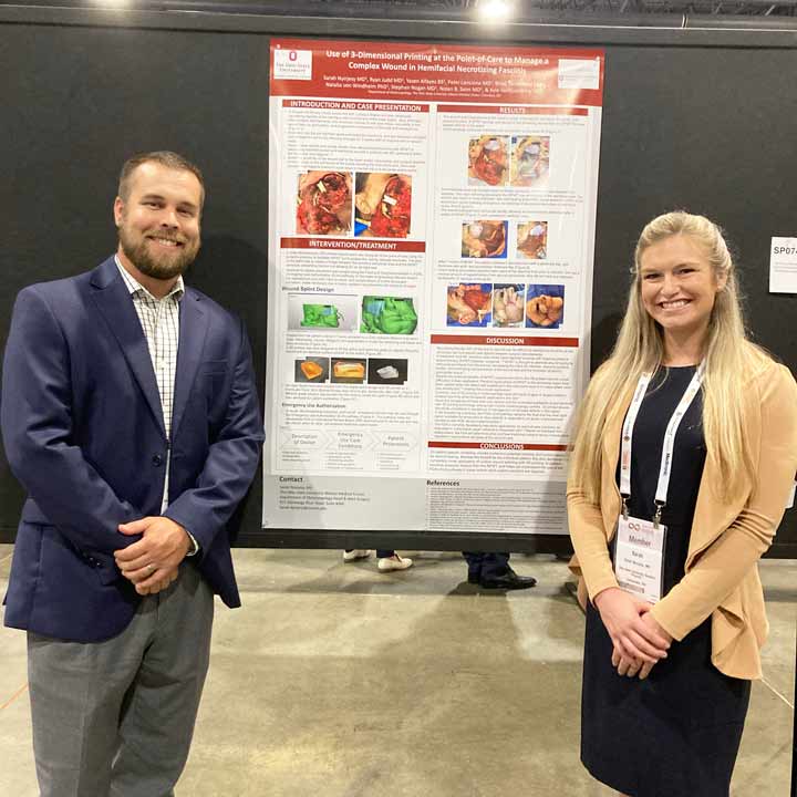
 Sarah Nyirjesy, MD, still has the crude drawing she and her team used to design a patient-specific facial splint on a 3D printer. The patient with rare hemifacial necrotizing fasciitis (NF) from Ludwig’s angina was deteriorating, even after a month of surgical debridements and rounds of IV antibiotics. Dr. Nyirjesy’s idea was to create a device to protect her patient’s eye while the team applied a negative pressure wound VAC to the rest of the facial wound.
With the customized 3D-printed silicone facial splint and wound VAC, the patient began to improve. It was a lightbulb moment that saved the woman’s life.
From idea to implementation, it took only 72 hours.
Treating a rare case of NF with debridement
One month earlier, Dr. Nyirjesy, at the time a third-year resident in the Department of Otolaryngology – Head and Neck Surgery at The Ohio State University Wexner Medical Center, was called to consult on an unusual infection in an oral surgery case. She guessed from the patient’s presentation that it was a rare case of NF – a rapidly progressive soft tissue infection with a high mortality rate. The infection can occur throughout the body, but its proximity to vital structures in the face and mediastinum made this case more concerning.
The 58-year-old female presented with NF of the neck and hemiface due to a tooth abscess. There was poor tissue vascularity in the wound bed and no evidence of healthy granulation tissue, despite a month of aggressive wound care, antibiotics and surgical debridements. Dr. Nyirjesy and her team were concerned about further breakdown toward the right orbit, mediastinum and pretracheal soft tissues — necessitating the patient remain intubated for this extended period of time.
A negative pressure wound VAC could have improved healing, but traction near the orbit would have caused vision loss.
With no other option, the patient remained intubated for a month. The plan was to continue treatment with antibiotics, aggressive wound care and operative debridements, but the prognosis wasn’t good.
That’s when Dr. Nyirjesy realized she could create a 3D-printed, patient-specific silicone wound splint for negative pressure wound therapy (NPWT). She and research engineering colleague Yazen Alfayez designed and printed the device from the patient’s CT scan to sit along the exact contour of the wound and serve as the contact point for the wound VAC, rather than on the eyelid.
They obtained approval using a Food and Drug Administration (FDA) pathway to manage rapidly changing, life-threatening clinical scenarios when no other existing therapy is viable. They completed the project over a weekend.
Dr. Nyirjesy was particularly interested in the potential use of 3D-printed medical solutions in otolaryngology and had joined the Translational Therapeutics Program at The Ohio State University Comprehensive Cancer Center – Arthur G. James Cancer Hospital and Richard J. Solove Research Institute (OSUCCC – James) under the mentorship of Kyle VanKoevering, MD, an otolaryngologist who specializes in treating skull base tumors and cancers. Through his teaching, she learned the basics of 3D printing and custom device design.
Dr. VanKoevering came to the OSUCCC – James in 2020 with a vision to build out an engineering research lab around 3D printing in medicine. Today, he’s the director of the Medical Modeling, Materials and Manufacturing Lab (M4 Lab). With his experience creating custom devices, he guided Dr. Nyirjesy through the splint design and helped her navigate the process to get device approval under the FDA’s Expanded Access for Medical Devices emergency use mechanism.
As a mentor, teaching the technical 3D lab skills to the next generation of physicians and then supporting a team to create a device that saves a patient’s life was precisely why Dr. VanKoevering led the lab’s development.
“One of the best things I get to be a part of as an academic surgeon is training the future generation,” he says. “People like Dr. Nyirjesy will take this field much farther than I ever will, with a mind like hers thinking outside of the box.”
The 3D wound splint saved the patient’s eye and life
After five days of splint-assisted vacuum therapy, the patient’s wound bed stabilized with no residual purulence. She developed healthy granulation tissue and sustained no eye or lower lid injury. The wound contracted with continued vacuum therapy to allow for safe tracheostomy placement, ventilator liberation and oral intake. The patient had hemifacial reconstruction with a myofascial pectoralis muscle flap and a paramedian forehead flap one month later. She was eventually decannulated and had excellent wound healing and periorbital function at a six-month follow-up appointment.
She and her family continually express gratitude for the customized care they received.
For Dr. Nyirjesy, who’s still early in her medical career, the case reminded her exactly why she became a physician.
“I got to know the patient once she woke up. She could talk to family and interact with them. It was amazing to see.”
The case for future applications of patient-specific medical devices
This case is the first reported use of a patient-specific, 3D-printed wound splint to allow the application of NPWT near the orbit while preventing orbital injury and vision loss.
“Her idea was brilliant. This was a natural extension of what we were doing, but it’s a radically different application,” Dr. VanKoevering says.
The wound splint highlights the potential for point-of-care medical devices to treat complex soft tissue defects from wounds near delicate, vital structures in the head and neck. This case is also an example of how physicians can successfully use the FDA’s investigational device exemption with patient and hospital consent.
“We have millions of medical devices on the market today, most of which come in one to three sizes,” Dr. VanKoevering says. “By making small tweaks in the manufacturing process with 3D printing, we can think about how to customize devices with better precision and performance. This opens up a whole new platform for products and completely changes how we treat patients.”
Together, these tools represent a powerful option for clinicians facing unprecedented medical conditions who may benefit from a customized solution delivered fast. They pave the path to do whatever it takes to maximize the potential outcomes for each patient.

2024 Year in Review
See how Ohio State is shaping the field of Otolaryngology – Head and Neck Surgery.

