First drugs ever approved for lupus nephritis

Ultrasonography is not yet widely adopted among U.S. nephrologists. However, its benefits – including portability and precision – have prompted kidney disease specialists at The Ohio State University Wexner Medical Center to use point-of-care ultrasound (POCUS) in the critical care setting and for patients on dialysis.
“Even though nephrologists are considered the experts in assessing and managing patients’ fluid status, traditional physical examinations, including assessing jugular venous pressure, auscultating for crackles or checking for pedal edema, are often inaccurate,” says Anil Agarwal, MD, professor of clinical medicine in Ohio State’s Division of Nephrology and director of the interventional nephrology program. “But new evidence confirms that ultrasonography can optimize fluid management among dialysis and other kidney disease patients.”
Results of the LUST trial (Lung Water by Ultra-Sound Guided Treatment to Prevent Death and Cardiovascular Complications in High Risk ESRD Patients with Cardiomyopathy), published by the Clinical Journal of the American Society of Nephrology in 2018, demonstrated that lung ultrasound was much better in assessing interstitial fluid in dialysis patients in comparison to lung auscultation. Leg edema was only 3% sensitive to detect volume overload, and adding auscultatory findings increased it to only 13%.
While other markers of volume status assessment have also evolved, a recent randomized controlled study found lung ultrasound to be superior to chest X-ray and NT-ProBNP. The study results, published in 2019 by the European Journal of Heart Failure, showed that lung ultrasound reduced diagnosis time to only 5 minutes – compared to 104 minutes using the traditional technique.
In Ohio State’s Division of Nephrology, clinicians have embraced POCUS as a tool for managing complicated patients. Its diverse uses include:
“In some cases, we are able to extend the assessment to focused cardiac imaging prior to formal echocardiogram,” Dr. Agarwal says. “This helps us diagnose critical conditions like cardiac tamponade or cardiogenic shock.”
Dr. Agarwal believes these emerging applications are only the tip of the iceberg. “With the advent of new portable devices,” he says, “POCUS is destined to become the next ‘stethoscope’ for modern nephrologists to manage complex patients.”
The following images highlight the value of ultrasonography:
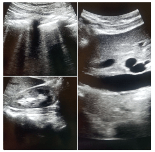
Shown are a patient’s lung ultrasound (top left corner), inferior vena cava (right) and kidney (bottom left). The lung image is consistent with so called “B-lines” (rays beaming from the top of the image to the bottom), the artifacts resulting from pulmonary edema. The inferior vena cava was dilated, confirming intravascular volume overload. The kidney appeared relatively normal. In contrast, the patient showed no physical signs of volume overload. The patient was treated for decompensated heart failure and recovered kidney function.