
The Ohio State University Wexner Medical Center expands treatment options for patients with COPD

 The ability to localize and sample small lung nodules has always been challenging to pulmonologists, even with the use of advanced techniques such as navigational bronchoscopy and radial endobronchial ultrasound. That’s one reason robotic-assisted bronchoscopy is the most innovative tool in the armamentarium of chest physicians for well over two decades.
The addition of a new robotic bronchoscopy platform to The Ohio State University Wexner Medical Center is helping physicians localize and gain access to the smallest lung lesions sooner and with greater ease.
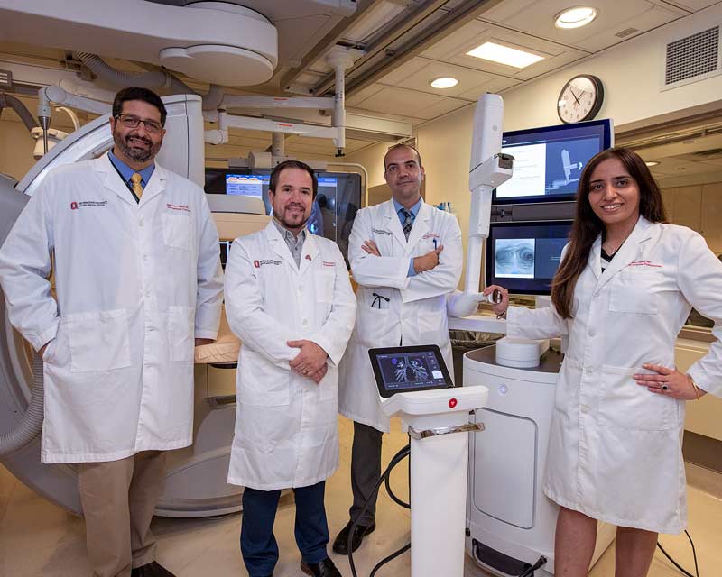
Alberto Revelo, MD, is an interventional pulmonologist who focuses on the diagnosis and management of pulmonary diseases, including lung cancers, using emerging technologies featuring minimally invasive methods. Together with his colleagues in the Division of Pulmonary, Critical Care and Sleep Medicine, he has used this new platform to help confirm more than 30 early-stage lung cancer diagnoses since August 2021.
The new platform features an ultra-thin, ultra-maneuverable catheter that provides access far into the lung periphery, and precision and articulation where standard bronchoscopy cannot reach.
“The robotic platform significantly improves our ability to localize and sample peripheral lung nodules,” Revelo says. “The robotic-assisted articulation significantly enhances the ability to negotiate multiple branch points in the tracheobronchial tree.”
Doctors at the Ohio State Wexner Medical Center have taken the technology a step further, combining it with intraoperative use of cone beam computed tomography (CBCT) to dramatically improve positional reference.
“We introduced cone beam CT to Ohio State in the fall of 2020, and now we frequently use it with the robotic bronchoscopy platform to better understand our position within the lungs as we biopsy the more difficult and smallest lung lesions that otherwise we would not get to,” Revelo says.
Cone beam CT uses a lower radiation dose, and it takes less than 10 seconds to obtain images intraoperatively, providing real-time information of the target lesion in relation to biopsy tools. The Ohio State Wexner Medical Center is one of the few centers nationwide offering these two technologies combined.
“Compared to other diagnostic methods, like CT-guided biopsy or transthoracic needle aspiration biopsy, the robotic bronchoscopy continues to be a lower-risk biopsy choice when used in conjunction with the current standard of care for mediastinal lymph node staging,” Revelo says. “This also allows for a single procedure approach to diagnosis and staging of thoracic malignancies. It’s a one-stop shop for both diagnosing and staging lung cancers. It’s important to offer patients more convenience and access to the latest technologies. That’s something we do very well here at Ohio State.”
The reliable localization within the central aspect of the lesion can potentially allow for bronchoscopic treatment of peripheral lung nodules in those patients who have early-stage lung cancers.
“Right now, the standard of care for early-stage lung cancers is surgery, or radiation therapy if surgery is contraindicated,” Revelo says. “We hope to one day offer some form of ablation therapy, like radiofrequency or microwave ablation, using the robotic bronchoscope and CBCT. Having the two technologies already is a huge step toward lung cancer diagnosis and therapeutics, and Ohio State is now at the forefront.”
Learn more about innovations in care and research from the Division of Pulmonary, Critical Care and Sleep Medicine.