
Swept-source OCT angiography reveals exquisite details of ocular structures

 A boon for advancing knowledge to improve outcomes for AMD, glaucoma and other eye diseases
For clinicians and researchers who are treating or studying age-related macular degeneration (AMD), diabetic retinopathy, glaucoma or other eye diseases, being able to see ocular structures is critical. Now, technology makes this visualization possible through noninvasive means.
Swept-source optical coherence tomography (OCT), introduced around 2012, is similar to spectral domain OCT. However, in swept-source OCT, the light source is “swept” through a range of wavelengths with each scan, and the temporal output of the detector is converted to spectral interference.
OCT angiography takes this technology one step further, reconstructing scans performed multiple times in a vertical plane into a single image shown on a horizontal plane — allowing noninvasive visualization of vascular structures in the retina and choroid without the use of contrast dye.
With the recent acquisition of this instrument, The Ohio State University Wexner Medical Center is applying this powerful technology to provide exquisitely detailed imaging data. This could lead to discoveries of potential biomarkers, enabling more effective evaluation, monitoring and treatment of AMD and many other eye conditions. With the acquisition of a specialized lens for the anterior segment in addition to the posterior lens, this technology will allow ophthalmology team members to investigate and explore structural details in healthy eyes and those in eye diseases with neovascularization, edema, ischemia or degeneration.
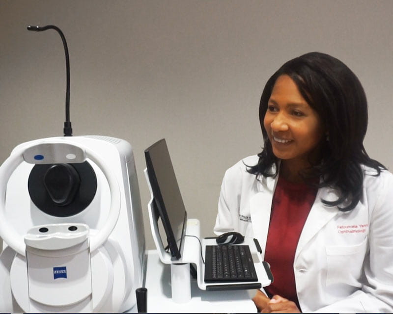
“This technology uses the movement of red blood cells to create high-resolution images of the retina vessels,” says Fatoumata Yanoga, MD, a retinal specialist and surgeon at Ohio State. “It also scans deeper, so we can see multiple layers of the eye in one scan, and it provides a wider view of the retina.”
AMD and diabetic retinopathy are two of Yanoga’s main clinical interests, so she’s hopeful about the potential for improving clinical outcomes and earlier detection.
“With AMD, there can be abnormal blood vessel formation in the choroid affecting retina and retinal pigment epithelium, and this technology will allow us to see those changes in detail and hopefully in earlier stages,” she says. “Since the angiography is noninvasive, these images will be easier on the patients and provide greater anatomical details for us. With my focus on AMD, that’s what I’m most excited about, but there are many other applications for this technology for both clinical and research purposes. With such exquisite, detailed imaging, we have the opportunity to discover structural biomarkers that may be more effective for earlier detection of disease and to track the progression of eye disease and monitor patients’ response to treatment. We will investigate whether these details provide new insights into disease mechanisms as well.”