improving patient care

 A 2024 pilot study at The Ohio State University Comprehensive Cancer Center – Arthur G. James Cancer Hospital and Richard J. Solove Research Institute (OSUCCC – James) demonstrated the efficacy of optical coherence tomography (OCT) imaging in helping ensure negative margins in small sections of soft tissue tumors. The researchers have now received an Ohio State presidential accelerator grant to take next steps in developing this novel application.
OCT uses near-infrared light to differentiate between tissue types. With its high axial resolution, OCT images can isolate transverse planes of tissues as thin as a micrometer, in 2- or 3-D.
A team of researchers from the veterinary, engineering and bioengineering colleges at Ohio State are collaborating with the OSUCCC – James to learn whether intraoperative OCT is equally discerning for examination of entire tumors, rather than just smaller segments.
Orthopedic oncologist Joel Mayerson, MD, director of the Division of Orthopaedic Oncology, medical director of the sarcoma program and associate chief medical officer of perioperative services for The James, is leading the medical side of the project.
“OCT has current applications in visualizing the retina and cardiovascular structures, for example, but only recently has it been applied to soft tissue tumors, and this is the first time that I’m aware of that anyone has combined AI with OCT to examine sarcomas in humans,” Dr. Mayerson said.
The project began with a conversation between Dr. Mayerson and Laura Selmic, BVETMED, MPH, the Teckie and Don Shackelford Chair in Canine Medicine at the College of Veterinary Medicine. Dr. Selmic was the principal investigator on a multi-site study using intraoperative OCT on tumor margin delineation in feline and canine patients. Specifically, she had been working with OCT to detect sarcoma margins in dogs, who are more susceptible to developing these tumors, when she approached Dr. Mayerson in 2022 to discuss her findings.
“Dr. Selmic was the mastermind behind all of this,” Dr. Mayerson says. “Together, we decided to collaborate to see if we could extrapolate the outcomes she was finding in dogs to humans.”
Sarcomas are highly aggressive bone or soft tissue tumors that can readily spread to other sites, with a high risk of local recurrence after surgical excision. Five-year survival rates range from 20-90%, depending on the stage and type of sarcoma.
“Bone sarcomas usually occur in kids and teenagers, and it is heartbreaking that about a third of these kids and teenagers are dying,” Dr. Mayerson says. “Survival rates are not much different today than they were 40 years ago because it’s rare, so there isn’t enough incentive for industry to develop new drugs. Because of this, we have had to figure out other ways to try to improve their survival rate.”
To this end, Dr. Mayerson helped develop the use of 3-D cutting guides for surgical planning in bone sarcomas, and helped design one of the implant systems for bone sarcomas as well as methods for reconstruction.
“All these are surgical advances to help patients be more functional,” he said. “Now, we’re trying to advance treatment from another angle.”
Determining negative margins intraoperatively helps avoid local recurrence and additional surgeries, reduces the risk of infection associated with additional surgery and allows for timely chemotherapy. Resection of any tumor is complicated, but assessing the entire tumor magnifies the challenge to make accurate margin assessments. Tumor shape and location can make margin assessment even more difficult at the time of surgery. New methods are needed to help the surgeon perform more precise tumor excisions by removing what is necessary but saving as much as possible to retain function of an extremity.
Looking at the MRIs, and drawing on his 25 years performing these surgeries, Dr. Mayerson says he generally aims for a one centimeter tissue cuff when resecting a tumor. Then a pathologist samples the margin every centimeter around the tumor looking for cancer cells
“Still, no matter how skilled and thorough the surgeon, it’s not possible to know for sure that you have gotten everything at the time of surgery with our current technology,” says Dr. Mayerson.
While OCT cannot identify microscopic cancer cells, it enhances gross evaluation by imaging in thin horizontal slices, and it can be used to scan segments of the area of concern for a close margin. Researchers at Ohio State are currently working to extend the scope of OCT to potentially the entire tumor, which may fill in the gaps not sampled by current techniques in pathology.
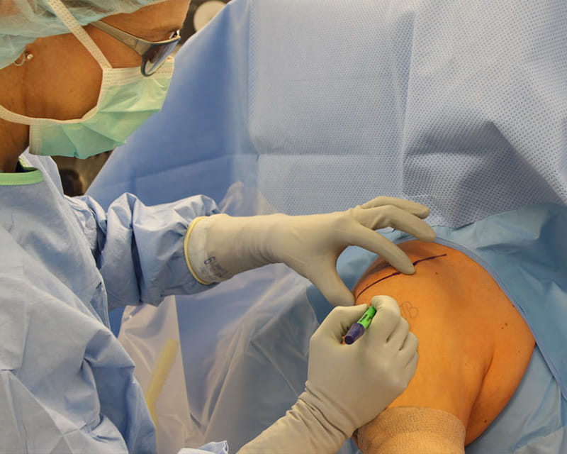
“You can take the probe, place it over the resected tumor, and it can tell the difference between fat, muscle, fascia and cancer, and do so in just one-to-two millimeter depths of tissue,” he says.
In their pilot study, the researchers took small segments of resected sarcomas from 20 patients and, in parallel with the pathologist’s inspection, compared OCT’s detection of positive margins at one centimeter intervals. They found a high correlation, with accurate tissue differentiation and margin assessment in the same 19 of the 20 patients.
Now, they are planning to test OCT over the surface of the entire tumor. This will require the development of AI code to see if computers can be taught to do this over a much larger surface area. The AI component is led by Ping Zhang, PhD, director of the Artificial Intelligence in Medicine Lab.
“The computer will hopefully give us a readout pretty quickly,” Dr. Mayerson says. “The pathologists will still follow their normal protocol, and the goal is to spend 10-to-15 minutes evaluating the tumor. This is to minimize the infection risks inherent in having an open incision site and decrease the chances of needing a second surgery if the initial margins are positive.”
Once the code is written, the plan is to do another human trial which, if successful, will serve as proof of concept. Then, the team’s hope is to secure a multi-institutional grant with a subsequent human trial, beginning sometime next year, and work toward utilization in human clinical medicine.
It took a confluence of different expertise and some unusual collaborations to even conceive of this project. Dr. Mayerson points to the renown of the Veterinary College of Medicine as the big starting advantage.
“It’s one of the top schools in the country,” Dr. Mayerson said. “There are 20 surgical oncologists in the veterinary world in the whole country, and three of them are here at Ohio State, treating over 100 dog osteosarcomas per year.”
This amalgam of four colleges and individuals’ expertise presents an opportunity to make one of the most significant changes in sarcoma outcomes in decades.
“If there’s a way that we can show that the margins are negative before the patient leaves the operating room, that’s an ideal world,” Mayerson said.
Importantly, Dr. Mayerson brings to the effort his longstanding work to build an academic multidisciplinary sarcoma program that improves survival.
When Dr. Mayerson first arrived at Ohio State, he was the only sacrcoma specialist.
“We are now one of the country’s largest programs, with 19 faculty members that take care of our sarcoma patients, including orthopedic oncologists, general surgical oncologists, medical oncologists, radiation oncologists, radiologists, pathologists and plastic surgeons,” he says.
“All of these people are involved in our weekly tumor board so we have multiple eyes on each patient,” he says. “In this group, the enthusiasm over having a more effective way to help clear margins is palpable.”