
 Under the leadership of Benjamin Segal, MD, chair of the Department of Neurology in The Ohio State University College of Medicine and director of the Neuroscience Research Institute at The Ohio State University Wexner Medical Center, faculty are embracing a deeply collaborative and integrated research culture — one that dissolves boundaries between disciplines and accelerates the movement of discoveries from laboratory benches to clinical application.
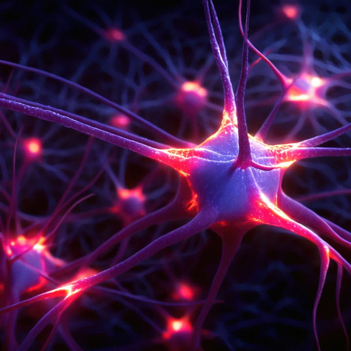
Within this environment, a new generation of neurology investigators is pursuing independent research programs that probe the cellular and molecular mechanisms underlying a spectrum of neurological disorders. Their efforts are laying the groundwork for innovative neuroprotective and pro-regenerative strategies that aim not only to prevent and mitigate neural injury but, ultimately, to repair disrupted neural circuits and restore lost neurological functions.
These three rising neuroscientists are conducting research that captures the breadth and promise of cutting-edge translational neurology – ranging from immune-neuronal interactions after spinal cord injury, to cutaneous regulation of sensory neurons in chemotherapy-induced neuropathy, to the role of lysosomal integrity in Parkinson’s disease. Together, their discoveries reveal new therapeutic pathways and advance the field toward a future in which clinically meaningful neuroprotection and neural regeneration are attainable goals rather than distant aspirations.
Reimagining the role of the immune system in neurological diseases
 Andrew Sas, MD, PhD, assistant professor of Neurology in the Ohio State College of Medicine, is overturning long-standing assumptions about innate immune cells. Neutrophils and monocytes – classically considered short-lived drivers of destructive inflammation in the central nervous system (CNS) – emerge in his work as unexpectedly potent contributors to neural repair.
Dr. Sas investigates how subsets of neutrophils communicate with neurons and CNS-resident glia, and how their beneficial signals might be sustained despite the fleeting lifespan of any single neutrophil.
As a postdoctoral fellow in Dr. Segal’s laboratory, Dr. Sas began by interrogating immune-neuronal interactions in a murine optic nerve crush model. Although sterile inflammation in the vitreous had been linked to axonal regrowth, the responsible immune cell type and molecular mediators were unknown. Dr. Sas and colleagues identified a previously unrecognized subset of immature neutrophils that secretes growth factors promoting retinal neuron survival and driving optic nerve regeneration.
“We asked very basic questions about how innate immune cells might interact with neurons in a beneficial way – what makes them protective and what enables them to support healing,” Dr. Sas explains. The team demonstrated that these specialized immature neutrophils directly stimulate axon regeneration through neurotrophic and neuroprotective factor production and, in parallel, modulate microglia and astrocytes to shape a repair-permissive microenvironment.
A critical advance came when the group developed a method to generate these pro-regenerative neutrophils in large numbers by polarizing bone marrow-derived precursors with a specific cytokine cocktail. When administered into the injured CNS, these engineered neutrophils drove robust axonal regeneration – not only in the optic nerve, but also in models of spinal cord injury.
“The ability to generate large numbers of these novel neutrophils makes them a potential cell therapy for patients who have suffered neurological injuries,” Dr. Sas says.
Perhaps most compelling, the team achieved axon regrowth in the spinal cords of mice reconstituted with human immune systems. Furthermore, they were able to regrow previously injured and stunted human neurons in vitro with polarized human immature neutrophils – underscoring the translational promise of this approach and its potential to one day reverse CNS injury.
As an independent investigator leading his own NIH-funded laboratory, Dr. Sas has expanded this work to reveal a broader, coordinated immune program of CNS repair. His team has shown that neuroprotective neutrophils do more than deliver direct trophic support – they also recruit and reprogram a second wave of infiltrating monocytes, pushing them toward reparative phenotypes and establishing a self-reinforcing cascade of immune-mediated healing.
In parallel, his group is dissecting how aging influences the capacity to generate neuroprotective, pro-regenerative innate immune cells. By defining the age-dependent hurdles in the polarization of reparative neutrophils and monocyte subsets, they aim to uncover therapeutic strategies that preserve or restore regenerative immunity across the lifespan.
The ultimate goal is to translate these findings into the first neuroprotective and restorative cell therapies for patients with CNS injury and neurodegenerative diseases.
“A major focus now is determining whether human cells can recapitulate the behavior of their counterparts in humanized mouse models – an essential step toward reproducible, scalable cell-based therapies,” he explains. “Close collaboration with subspecialty neurologists is critical to ensure the science remains aligned with real clinical needs.”
Looking ahead, Dr. Sas expects future applications for a wide variety of neurological injuries, including stroke, traumatic brain and spinal cord injury, and even autoimmune conditions such as multiple sclerosis.
Crosstalk between skin cells as key to post-chemotherapy neuropathic pain
 While neuropathic pain is classically conceptualized as a neuronal problem, Grace Ji-eun Shin, PhD, assistant professor of Neurology in the College of Medicine, argues that the surrounding skin microenvironment is a critical – and profoundly understudied – regulator of sensory neuron health, particularly in the context of chemotherapy-induced peripheral neuropathy (CIPN).
Dr. Shin says that the majority of people who receive chemotherapy develop CIPN, presenting with numbness, tingling or severe pain in their limbs. Some patients recover, while others develop chronic conditions that can last a lifetime. With collaborations spanning neurology, dermatology, oncology and pharmacy, Dr. Shin’s research seeks to define how skin-resident cell types – including keratinocytes, macrophages and fibroblasts – interact with sensory neurons to determine whether damaged axons regenerate or remain chronically dysfunctional.
This line of research required her team to recapitulate the cutaneous microenvironment in drosophila, mouse and human cell models, enabling precise dissection of immune, stromal and neuronal interactions during exposure to chemotherapeutic agents. By generating personalized hybrid cultures of patient-derived immune cells and sensory neurons (the latter generated from induced pluripotent stem cells), her team is identifying master regulators of repair resistance – potential therapeutic targets for skewing the skin environment toward neural regeneration.
“What we are hypothesizing is that by targeting the orchestrator, the impact will propagate to other cells,” Dr. Shin says.
Dr. Shin collaborates closely with neurologists caring for people with CIPN at Ohio State to obtain skin biopsies from well-characterized patients. By dissecting the interactions between sensory neurons and other cell types in the biopsied tissues, Dr. Shin is aligning cellular and molecular discoveries with clinical phenotypes, enabling future stratification of patients most likely to benefit from targeted interventions. She’s also working with pharmacologists and collaborators at Columbia University to identify repurposed therapeutics capable of preventing or reversing chemotherapy-induced neurotoxicity.
“My dream would be that drugs used for other neurological or dermatological conditions could be repurposed to treat CIPN in oncology and neurology clinics everywhere,” Dr. Shin says.
Her group is now extending this model to other forms of peripheral neuropathy, including traumatic spinal nerve injury, broadening the translational reach of her research.
Elucidating how lysosomes fail in Parkinson’s disease
Abnormal aggregation of misfolded proteins is a common characteristic of many neurodegenerative disorders, including Parkinson’s disease (PD), Alzheimer’s disease and amyotrophic lateral sclerosis (ALS). Under normal conditions, lysosomes serve as the cell’s “street sweepers,” degrading misfolded proteins and clearing protein aggregates. In PD, however, lysosomal dysfunction accelerates aggregate buildup and ultimately contributes to neuronal degeneration.
 Luis Bonet-Ponce, PhD, an assistant professor of Neurology in the College of Medicine, is investigating how lysosome membranes become damaged in PD and how this process links genetic mutations to downstream cellular dysfunction.
“When you culture neurons with aggregates from these diseases, the aggregates invariably end up in lysosomes,” Dr. Bonet-Ponce explains. “When lysosomal membranes are damaged, proteins can’t be broken down and discarded. We see this pathway of membrane injury across virtually every neurodegenerative disease, and even in cancer, muscle and lung disease and infection.”
His earlier work at the NIH demonstrated that leucine-rich repeat kinase 2 (LRRK2), a kinase whose gain-of-function mutations cause familial PD (which constitute about 20% of cases) and increase the risk of sporadic PD, is actively recruited to damaged lysosomes. Excess LRRK2 activity impairs the cell’s lysosomal repair machinery, worsening membrane instability.
“We know that protein aggregates must be degraded by lysosomes, including debris phagocytosed by microglia and macrophages,” he says. “And lysosomal membrane damage is a consistent feature of neurodegeneration. So the key question became: how do LRRK2 mutations derail lysosomal homeostasis?”
Dr. Bonet-Ponce’s lab has recently identified a repair-disrupting mechanism they call LYTL (LYsosomal Tubulation/sorting driven by LRRK2). In this process, lysosomes exposed to hyperactive LRRK2 form tubules that bud off as mobile vesicles. These vesicles then fuse with healthy, intact lysosomes and spread membrane damage, creating a cascade of dysfunction throughout the cell.
The team is also studying how mutations in other lysosomal membrane proteins associated with PD – transmembrane protein 175 (TMEM175) and ATP13A2 – alter ion transport and metabolic flux, further compromising proteostasis.
“Lysosomal membrane damage appears to be a convergent feature across neurodegenerative diseases,” Dr. Bonet-Ponce explains. “Understanding how specific mutations disrupt this system allows us to identify new therapeutic targets – and potentially repurpose drugs for genetically defined subtypes of disease.”
He adds a note of caution: treatments will likely need to be tailored to individual genetic backgrounds.
“We need to study them, mutation by mutation. Then, I think it is reasonable to expect that we’ll be able to repurpose drugs to treat many of these variants,” he says.
A convergence toward repair-focused neuroscience
What unites these diverse research programs is a shared commitment to translating a deeper understanding of disease mechanisms into therapies that actively promote repair in the damaged nervous system. Whether harnessing immune cells to drive axon regeneration, decoding how the skin shapes sensory neuron health or restoring lysosomal function in neurodegenerative disorders, these investigators have a common goal: redefining what’s possible in translational neuroscience.
Dr. Segal notes that this culture of integration – spanning the full arc from bench to bedside and from neurons to immune and skin cells – creates the conditions for discoveries that once felt out of reach.
Together, Ohio State’s rising neurologists are helping build a future in which neuroprotection and even neural regeneration ascend from aspiration to attainable reality.