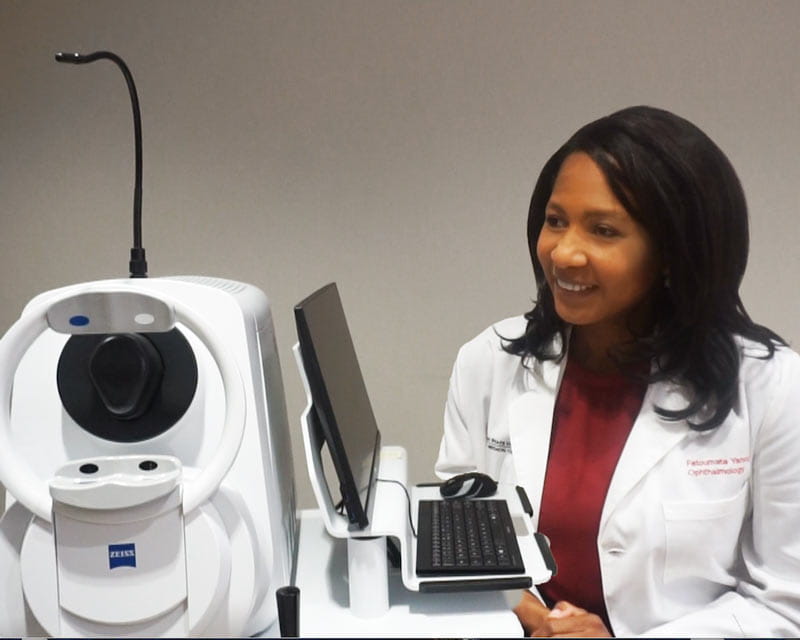
Swept-source OCT angiography reveals exquisite details of ocular structures
— Greater understanding of the mechanisms behind ocular diseases and what causes them to progress could potentially improve ophthalmology care and lead to better visual outcomes for patients. Research led by Cynthia Roberts, PhD, of The Ohio State University Wexner Medical Center may be on the verge of doing both.
An R01 grant Dr. Roberts received in 2016 to study corneal biomechanics was renewed in July for nearly $1.7 million. It allows her to build on earlier findings and develop biomechanically driven risk models with a longitudinal component for keratoconus, diabetes and glaucoma.
As expected, Dr. Roberts’ earlier work revealed that in healthy corneas, stress distribution is dominated by corneal thickness. But things differ in keratoconus, and stress distribution becomes dominated by curvature.
“I have proposed that there’s a biomechanical cycle of decompensation driven by asymmetry of properties,” Dr. Roberts says. “So the focal area of pathology is weakened, but the rest of the cornea is not. The weak area deforms more than the rest, so it stretches and becomes thinner. As it thins, the corneal response is to focally increase curvature, because greater curvature has less stress.”
The “corneal contribution to stress” metric can be calculated with curvature and thickness information simultaneously incorporated in tomography and used to predict advancing keratoconus. Collaboration with researchers from Maastricht University in The Netherlands, who had longitudinal data, has already shown that baseline difference in maximum and minimum stress significantly predicts five-year progression in keratoconus.
Insurance companies typically require documentation of progression before covering corneal cross-linking. But in some cases, by waiting for documentation, the patient is no longer eligible for the procedure due to increased severity of disease. Predicting progression from a single stress map could be a game changer that leads to earlier treatment and spares patients from more invasive corneal transplants.
Andrew Hendershot, MD, an ophthalmologist who specializes in cornea and external eye disease, is collaborating with Dr. Roberts on this research.
While Dr. Roberts’ original grant focused on corneal biomechanics, her work led to important discoveries about the sclera and diabetic retinopathy.
Using biomechanical assessment devices that included an air puff to perturb the cornea, she measured corneal stiffness in two groups: people who had diabetes but didn’t have diabetic retinopathy and those who had diabetes with diabetic retinopathy — a parallel protocol funded by the Ohio Lions Eye Research Foundation.
Evidence shows that with hyperglycemia, accumulation of advanced glycation end products (AGEs) leads to nonenzymatic cross-linking and subsequent stiffening of the cornea. Dr. Roberts’ research found it’s not just the cornea that stiffens. In people with diabetic retinopathy, there was also a stiffer scleral response.
“When an air puff is applied to the cornea, the cornea becomes concave,” she says. “As it becomes concave, it displaces fluid. That fluid has to go somewhere, so the sclera expands. If the sclera is stiff, corneal motion is limited, which might be misinterpreted as a stiffer cornea, not a stiffer sclera.”
Realizing that scleral stiffness was impacted was unexpected and, initially, surprisingly hard to explain. In discussion with retinal specialists, Dr. Roberts understood the real issue.
“If the cornea experiences stiffening due to AGEs, then, of course, the sclera does too,” she says. “It’s just no one has looked at the sclera.”
Dr. Roberts believes a stiffer sclera is a cumulative indication of long-term hyperglycemia, and with longitudinal follow-up in people with diabetes, she’ll use the biomarker together with hemoglobin A1c to establish a risk model of developing diabetic retinopathy. If found to be a predictor of the disease, measuring scleral stiffness may be incorporated into diabetic patients’ annual eye exams and efforts to improve glucose control, if needed, can be further emphasized.
Ohio State collaborators on this research include Matthew Ohr, MD, Alan Letson, MD, and Jeff Pan, PhD.
The 2002 Ocular Hypertension Treatment Study (OHTS) demonstrated that thin corneas in those who have ocular hypertension leads to an increased risk of glaucoma.
Based on her earlier findings on the relationship between elastic and viscoelastic parameters in glaucoma and ocular hypertension, including the sclera’s role in dissipating energy from a corneal deformation, Dr. Roberts is planning to add corneal and scleral stiffness and corneal hysteresis to the existing risk model from OHTS. Since these measurements weren’t possible when OHTS was conducted, they may offer a potentially improved indication of risk for glaucoma conversion.
Gloria Fleming, MD, and Phil Yuhas, OD, PhD, collaborate with Dr. Roberts on glaucoma research.