

What is a cardiac MRI?
A cardiac MRI is a safe, noninvasive test that shows images of the heart and large blood vessels from any angle. A cardiac MRI is a powerful tool used to various forms of heart disease to help manage conditions such as coronary artery disease, heart failure, arrhythmias, valve diseases, heart inflammation, heart tumors and congenital heart defects. Along with cardiac stress tests (exercise stress test, echo stress test, nuclear stress test) and cardiac CT scans, cardiac MRI is a powerful complementary tool that helps to provide the most complete cardiac information to your physician.
Why choose Ohio State for your cardiac MRI
We have central Ohio’s most advanced cardiac diagnostic testing capabilities operated by heart specialists — empowering us to give you the best and most comprehensive cardiac care. Every year our team performs over 2,500 cardiac MRIs.
The Ohio State University Wexner Medical Center is one of only a few academic health centers in the country with three cardiac MRI scanners allowing us to image patients with a variety of conditions including those with implanted hardware or who may be claustrophobic or too large for traditional MRI scanners.
Even more important than our technology is our team of cardiology experts who work closely with your referring doctor to ensure the most accurate and comprehensive results of your tests.
Cardiac MRI services at Ohio State
We offer multiple types of cardiac MRI services to provide the needed analysis of your heart.
- Cardiac MRI (CMR) – This is the most detailed and comprehensive non-invasive evaluation of your heart that can help diagnose and guide treatment for a wide range of heart conditions, including cardiomyopathy, heart valve diseases and congenital heart diseases
- Stress cardiac MRI – using adenosine and exercise, we offer comprehensive evaluation for coronary artery disease and cardiomyopathy
- Cardiac MRI in the presence of implantable conditional or non-conditional devices
- Cardiac MRI for those who can't fit into conventional scanners due to body size or claustrophobia
- MRI angiography (MRA) – a tool used to diagnose and monitor vascular diseases
What to expect during a cardiac MRI
Preparing for your procedure
Avoid caffeinated foods and drinks, as well as smoking for at least 12 hours before your test. In addition, do not eat or drink anything, other than small sips of water for daily medication, for three hours before your test. Your physician may provide you with oral medication to take prior to your cardiac MRI to help you relax, especially if anxiety or claustrophobia are issues.
You will be asked to remove all metal objects from your pockets and body, including jewelry, coins, keys, hearing aids, cellphone and watch. Notify the technician if you could be pregnant, if you have a cardiac pacemaker or defibrillator, any type of implanted device, metal shrapnel or fragments inside your body, or if you are claustrophobic or become anxious in confined spaces.
Once the technologist is ready to begin, a contrast IV will be started in your arm to provide a clearer MRI picture.
During your procedure
Follow the technologist’s instructions closely and make sure to hold completely still. Your cardiac MRI should last between 30 and 60 minutes, so please be patient.
You will be placed on a table that rolls into a large, tube-shaped machine. Your entire body will be inside the tube for the entire test. The technologist will communicate with you during the test and give you instructions on holding your breath or holding a certain position. It is essential that you stay as still as possible during the test so the pictures are clear. The machine also makes a lot of loud knocking noises during the test — this is completely normal. You will have headphones on to help you communicate with the technologists, and you can opt to have music playing during the study.
After your procedure
If you received a sedative to help you relax during the test, you need to have an adult present to take you home. Drink plenty of noncaffeinated liquids after the test to flush the contrast medicine out of your body.
Once you are free to go, you can resume normal daily activities. A report of the test will be sent to your physician, who will contact you to discuss your results. If you feel something is not right after your test, please call your physician right away.
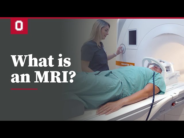
What is an MRI?
In this video, our imaging technologist explains an MRI (magnetic resonance imaging), what’s used for and what you can expect during this type of imaging exam.

Karolina Zareba, MD, a cardiologist at The Ohio State University Wexner Medical Center, describes a cardiac MRI test and what you can expect as this test is performed.
Get an estimate for this and other services
Get an instant estimate for your out-of-pocket costs. Learn more.
Login to MyChart
As a MyChart user, you can pre-populate an estimate with your existing insurance information, receive personalized estimates and save your estimates for later.
Don't have a MyChart account? Create an account