
Prenatal Ultrasound
With ultrasounds and our advanced imaging techniques, we can monitor your baby’s growth and development, providing you with peace of mind during your pregnancy.

 What is a prenatal ultrasound?
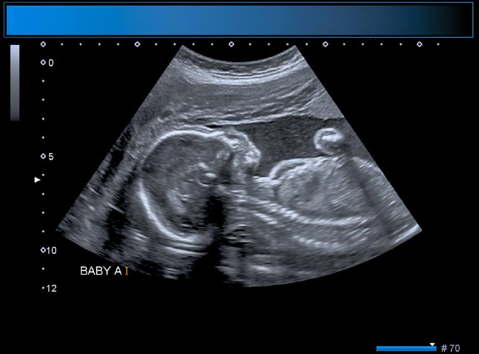
An ultrasound is one of the most commonly used prenatal tests during pregnancy that gives your provider more information about your baby's health. During a prenatal ultrasound, sound waves produce images of the baby inside the uterus, allowing you and your provider to see the baby’s organs and how the baby is developing. Measurements of the baby may be taken during an ultrasound to check the baby’s growth.
Most people can expect to receive one ultrasound during their pregnancy. However, if your pregnancy is high-risk, you may receive more than one ultrasound while receiving expert care in our award-winning Maternal Fetal Medicine Program.
The vast majority of the 30,000 obstetrical ultrasounds done by our obstetrics providers at The Ohio State University Wexner Medical Center in Columbus, Ohio, are the 2D traditional version, as that’s the only test that allows us to see the baby’s organs. While the 3D and 4D versions can produce cute pictures of your baby in the womb, they’re not done routinely. They’re only used if needed to provide specific clinical information, such as any facial anomalies.
How early will a pregnancy show up on an ultrasound?
Here’s what an early pregnancy might look like on an ultrasound:
- At five to six weeks: A pregnancy can usually be seen on ultrasound around five to six weeks after your last menstrual period. At this stage, your provider can often detect the gestational sac inside the uterus and the first look at the baby.
- By about six to seven weeks: A fetal heartbeat can usually be detected on a transvaginal ultrasound, which provides a closer and more detailed view in early pregnancy.
- Between eight and 12 weeks: The first official ultrasound is usually performed at this time. This early scan confirms the pregnancy, estimates your due date, checks for the baby’s heartbeat and determines whether you’re carrying one baby or multiples.
If you have a history of miscarriage, ectopic pregnancy or other early pregnancy concerns, your provider may schedule an ultrasound sooner to confirm that the pregnancy is progressing normally.
When an ultrasound identifies a pregnancy loss
Sometimes, an ultrasound reveals the loss of a pregnancy. Whether this occurs early or late in pregnancy, the loss of a baby can bring profound grief. If you’ve experienced a miscarriage, your care team can help you understand what happened, guide your physical recovery and connect you with counseling, support groups or spiritual care services.
Why is an ultrasound performed?
 In addition to giving expectant parents a chance to glimpse their growing baby, ultrasounds have many clinical uses during pregnancy. Ultrasounds not only measure the baby's size but also help identify issues and diagnose medical conditions.
An ultrasound might be performed to:
- Determine how many weeks pregnant you are
- Check if you’re carrying more than one baby
- Measure the baby’s size
- Look for birth defects
- Examine the baby’s heart rate
- Screen for risk of a premature delivery
Your provider may recommend additional screening or diagnostic prenatal tests as part of an ultrasound or to follow up on any abnormal results. These tests can include an amniocentesis or chorionic villus sampling.
Types of ultrasound
Generally, ultrasounds are done either transvaginally or abdominally. Both methods use safe, high-frequency sound waves to create images of your baby and uterus. The type of ultrasound you need depends on your pregnancy stage and what your provider needs to evaluate.
Transvaginal ultrasound (TVS)
This ultrasound is typically done earlier in pregnancy by inserting a slender wand, or transducer, into the vagina.
In early pregnancy, you can expect to see detailed images of the gestational sac, yolk sac and developing baby, as well as early signs of the placenta and fetal heartbeat. This type of ultrasound helps confirm the pregnancy’s location, estimate how far along you are and check for multiple fetuses.
Later in pregnancy during the second trimester, TVS is used to check the cervix length, which is a test to look at risk for premature birth, or sometimes to evaluate the location of the placenta.
Abdominal ultrasound
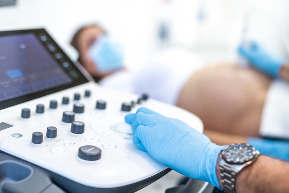
When the baby grows a bit bigger, an ultrasound can be performed by simply placing a transducer on the belly. As the pregnancy progresses and the baby grows, your provider will use an abdominal ultrasound. This method involves spreading a thin layer of gel on your belly and moving the transducer across the surface to create images.
Abdominal ultrasounds are most common in the second and third trimesters and are used to monitor your baby’s growth, anatomy, position, movement and heartbeat. They also allow your provider to assess the placenta, amniotic fluid levels and uterine structures. This type of ultrasound is entirely noninvasive and painless.
Common ultrasound examinations
Here are some prenatal exams that require an ultrasound:
- Anatomy scan: About halfway through your pregnancy (18-20 weeks), you’ll have a detailed ultrasound that looks at the baby’s organs, size and weight and overall health.
- Cervical length measurement: Measuring the cervix can help assess the risk of premature birth. This is usually done at the same time as the anatomy scan.
- Nuchal translucency: This screening test for detecting chromosomal disorders is performed between 11 and 13 weeks. The ultrasound measures the collection of fluid under the baby’s skin at the back of the neck.
- Doppler examination: This technique can be used to evaluate blood flow within your pelvic vessels and the baby’s blood vessels, including the umbilical cord. It’s only done in some high-risk pregnancies.
- Fetal echocardiogram: A special ultrasound that looks at whether the heart looks normal and works correctly, this test is typically done after 20 weeks for women who have a higher risk of having a child with a birth defect involving the heart. It’s done in partnership with Nationwide Children’s Hospital.
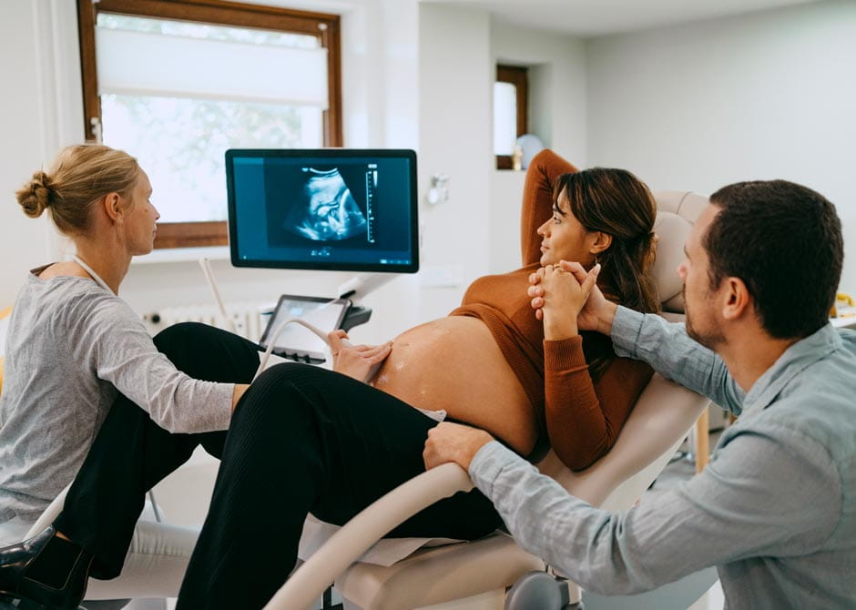
What to expect during an ultrasound
 This prenatal test will be performed by one of our certified sonographers in imaging or at the hospital. There are some key differences depending on what type of ultrasound you are receiving.
Transvaginal ultrasound
You’ll lie on your back, and a wand will be inserted into the vagina while your feet are in stirrups. You might feel some pressure from the wand but not pain. This typically lasts a few minutes.
Transabdominal ultrasound
This ultrasound is also performed while you lie on your back. Sometimes you may be asked to roll on your side. The sonographer will apply gel to your belly to help sound waves move more easily to your uterus. Moving the transducer across your belly creates images of the baby.
This ultrasound typically lets you see your baby’s face, hands, movements and biological sex for the first time. It’s important to remember that this is a diagnostic procedure, and our priority is to obtain diagnostic images to help our physicians determine the health of your baby. You’re not permitted to record during the ultrasound examination, and we’ll send you a link to electronically view your images after the exam is complete. We can’t guarantee that you will see the baby’s face, nor do we routinely provide 3D or 4D images. Again, our focus is on the baby’s health.
Results of an ultrasound
Video screens are positioned in the ultrasound room so that you’ll be able to view the images as the test is happening. After the ultrasound, you will be sent digital images of your baby.
You will need to wait for a provider to review and then relay the results.
Risks of an ultrasound
There are no known risks to you or your baby from an ultrasound. Unlike X-rays, ultrasounds use sound waves, not radiation, to create images.
That said, experts recommend avoiding unnecessary ultrasounds offered in private boutiques or nonmedical settings. These sessions may use equipment for longer periods or by technicians who aren’t certified in obstetric imaging.
How many times is an ultrasound done during pregnancy?
Most people have one or two ultrasounds during a healthy pregnancy. The two most common instances where you might receive an ultrasound include:
- First-trimester ultrasound (around eight to 12 weeks): Confirms the pregnancy, estimates your due date and checks for the baby’s heartbeat, number of fetuses and basic anatomy.
- Second-trimester ultrasound (the anatomy scan, around 18 to 22 weeks): A detailed exam that assesses your baby’s growth, organs, placenta and amniotic fluid. This is the screening that often reveals your baby’s sex if you’d like to know.
Your provider may recommend additional ultrasounds if you have a high-risk pregnancy, twins or multiples, or certain medical conditions, such as gestational diabetes or high blood pressure.
Why choose Ohio State for your ultrasound and prenatal care?
Here at the Ohio State Wexner Medical Center, all of our sonographers are certified. This ensures the best images of your baby during every ultrasound. This can give you peace of mind that your pregnancy is progressing as expected. However, if a potential problem is identified, our expert Maternal Fetal Medicine team can quickly provide an accurate diagnosis and offer unmatched support and care throughout the rest of your pregnancy.
If your pregnancy is considered high-risk for any reason, here are some of the benefits of being treated at the Ohio State Wexner Medical Center:
- We have specialized Maternal Fetal Medicine physicians, nurses and genetic counselors who care for high-risk pregnancies. For example, we provide prenatal care and delivery planning for all babies identified to have fetal anomalies or for pregnancies with red blood cell antibodies that require transfusion.
- Our team collaborates closely with Nationwide Children’s Hospital to ensure the continuity of care and delivery planning for high-risk babies.
- We have more than a decade of experience performing early fetal echocardiograms.
- Our licensed and board-certified team of genetic counselors will review your family history, assess your risks, explain your testing options, help you understand your test results and support you through your pregnancy whenever you have questions or concerns.
- Our new, state-of-the-art University Hospital and maternity center, opening in early spring of 2026, features stunning private rooms and the finest maternity amenities in central Ohio.
Get an estimate for this and other services
Get an instant estimate for your out-of-pocket costs. Learn more.
Login to MyChart
As a MyChart user, you can pre-populate an estimate with your existing insurance information, receive personalized estimates and save your estimates for later.
Don't have a MyChart account? Create an account