Building the bridge from discovery to treatment
Translational research is a key component of understanding and treating medical challenges. By working across the spectrum, from small animals to human subjects, this area of research transforms laboratory observations into actual treatments for the public.Andrew Sas, MD, PhD

Role of inflammation and immunomodulation in promoting recovery after traumatic neuronal injury.
Dr. Sas’s lab studies inflammatory responses to traumatic injury of the central nervous system (CNS). Among the immune cells that accumulate at sites of traumatic neuron injury, there is a subpopulation of myeloid cells that promote neuronal survival and regeneration of injured axons. Dr. Sas and colleagues are investigating the migratory pathways that drive the recruitment of pro-regenerative myeloid cells to the CNS. Based on an increased understanding of these migratory pathways, as well as the interaction between pro-regenerative myeloid cells and injured neurons, they’re working to develop novel immunotherapies that enhance the recovery of injured neurons in the eye, optic nerve, brain and spinal cord following trauma.
Specific Grants Awarded: K08
View Dr. Sas's profile Sas Lab
Stephen J. Kolb, MD, PhD

Genetic and molecular basis of motor neuron selectivity in neuromuscular diseases including amyotrophic lateral sclerosis and spinal muscular atrophy.
Dr. Kolb’s research centers on recent advances in the genetics of amyotrophic lateral sclerosis (ALS) and the emergence of therapeutic gene therapies, which has resulted in increasing optimism that there’s hope for meaningful disease-modifying therapies in genetic forms of ALS. Whole genome/exome sequencing and genome-wide association studies recently revealed that ALS-associated mutations in the C-terminal domain of the kinesin KIF5A. Dr. Kolb’s lab has created a novel mouse model of Kif5a ALS, with which the scientists will characterize the behavioral, electrophysiological and pathological changes as a first step to understand the functional consequences of KIF5A ALS-associated mutations and to create a tool for future preclinical and basic science studies.
Specific Grants Awarded: U24 , R03
View Dr. Kolb's profile Kolb LabBenjamin Segal, MD
 A novel inflammatory cell with neuroprotective and neuroregenerative properties
Chronic or progressive disability in individuals with central nervous system (CNS) injury is secondary, in large part, to the limited regenerative capacity of nerve fibers in the optic nerve, brain, and spinal cord. The long term goal of this project is to develop novel therapeutic interventions that overcome barriers to CNS repair and promote neuronal survival and axonal regrowth, thereby mitigating, or even reversing, neurological disability. The Segal lab has discovered a novel type of immune cell that migrates to sites of CNS injury, rescues neurons from cell death, and stimulates the regrowth of severed nerve fibers. The lab is currently investigating the lineage, phenotypic characteristics, and mechanism of action of this leukocyte subset. Ultimately, this research may lead to the development of novel autologous cell therapies, and/ or immunomodulatory drugs, that induce CNS repair and drive the recovery of lost neurological functions, in individuals with a range of neurological disorders including traumatic brain and spinal cord injury, multiple sclerosis, optic neuropathy, stroke and ALS.
Arginase-1 and iNOS expressing CNS myeloid cell sunsets in EAE and MS
Multiple sclerosis (MS), an inflammatory demyelinating disease of the central nervous system (CNS), is the most common cause of non-traumatic neurological disability in young adults in the Western Hemisphere. Significant progress has been made in the development of disease modifying therapies (DMT) that decrease the frequency of clinical MS relapses by blocking or depleting pathogenic lymphocytes. However, none of the approved DMT are curative, and none are effective in all patients. There are no treatments that slow, or reverse, progressive forms of MS. Myeloid cells, including macrophages, dendritic cells and microglia, comprise the majority of the immune cells that accumulate in the central nervous system (CNS) during Multiple Sclerosis (MS). Distinct CNS-infiltrating myeloid cell subsets have been implicated in damage or repair. Members of the Segal lab are studying the diversity, plasticity, biological properties, and function of CNS myeloid cells during exacerbations and remissions of experimental autoimmune encephalomyelitis (EAE), a widely used animal model of MS. With collaborators, they are also characterizing myeloid cell subsets in postmortem MS tissue. This research may lead to the identification of novel myeloid biomarkers and therapeutic targets for the management of individuals with relapsing and progressive forms of MS who do not respond to currently available treatments.
Specific Grants Awarded: R01, R01, R01
View Dr. Segal's profile Segal Lab
Billur Akkaya, MD, DPhil
 Deciphering the antigen mediated interactions and suppression mechanisms of human Tregs
This project aims to implement a novel pipeline to reveal the specificities of dominant autoreactive effector T cell and Treg clones derived from type 1 diabetes mellitus and multiple sclerosis patients. The overarching goal of this study is to devise new antigen-targeted adoptive Treg therapies for autoimmune diseases.
Discovering the molecular machinery underlying Treg interactions and Treg-mediated suppression
This project aims to elucidate the signals provided to Tregs via antigen mediated contacts with dendritic cells. We compare Tregs and effector T cells using conventional techniques such as phosphoflow and western blot while also performing an unbiased analysis of phosphoproteome, to decipher the pathways underlying antigen- MHC Class II capture and antigen-specific suppression. Overall, this project will uncover the molecular basis of antigen-specific suppression performed by Tregs and identify key molecules that can potentially be targeted to fine-tune the Treg activity in autoimmunity and cancer.
Determining the antigen-presentation capability of antigen-specific Tregs post- primary immune synapse
In this project we focus on the events after Treg-APC synapse and investigate whether capturing antigen- MHC Class II (pMHCII) equips Tregs with the unique ability to target helper T cells during the physical absence of dendritic cells. We perform live confocal and intravital two-photon microscopy imaging as well as flow cytometry 1) to quantify direct contact between Tregs and helper T cells post-pMHCII acquisition 2) to determine modes of paracrine communication. Altogether, the findings from this project will describe how the Treg-T cell interactions shape peripheral tolerance and what mechanisms additional to pMHCII depletion are in place to prevent autoimmunity and also to promote tumor development.
View Dr. Akkaya's profile Akkaya Lab
Bogdan Beirowski, MD, PhD
 Protecting axons through bioenergetics and glia
Axons are a particularly vulnerable component of neural circuits that degenerate in early stages of many debilitating neurodegenerative conditions such as Alzheimer’s and Parkinson’s disease, Multiple sclerosis, and various neuromuscular disorders. Therefore, the preservation of axons is an important therapeutic target. Dr. Beirowski’s laboratory pursues to elucidate the molecular and cellular mechanisms of axon degeneration and possible ways to counteract it. To develop novel approaches for axon protection the lab aims to capitalize on recent discoveries implicating neuronal and glial bioenergetics and their dynamic interplay.
Specific grants awarded: R01, R01
View Dr. Beirowski's profile Beirowski Lab
Elisabetta Babetto, PhD
 Energizing axons to combat neurodegeneration
An etiological commonality among many neurodegenerative disorders is the early degeneration of axons, which prevents neurons communicating with each other. Dr. Babetto’s research has contributed to the now widely established model that axons possess their own auto-destruction program that pivots on axonal energy metabolism. Her current focus builds on her discovery that the energetic depletion of perturbed axons can be antagonized by their adjacent glial cells though a mechanism called “axoglial metabolic coupling”. This discovery unveils new targets for developing therapies to slow or block axon degeneration in a variety of neurological conditions.
View Dr. Babetto's profile Babetto Lab
Cole Harrington MD, PhD
 Oligodendroglial heterogeneity and functional diversity in the context of an autoimmune inflammatory environment and aging
The primary goal of Dr. Harrington’s lab is understanding how oligodendroglia, the cells that make myelin and wrap axons, are altered in an inflammatory environment and during aging. In multiple sclerosis the autoimmune response results in damage to myelin sheaths or demyelination and loss of oligodendroglia. Oligodendroglia are necessary for restoring myelin sheaths and preventing axonal loss and neurodegeneration in MS lesions. In autoimmune inflammatory mouse models and human MS tissue oligodendroglia assume a variety of transcriptional phenotypes that likely influence their ability to proliferate, migrate, express mature myelin proteins and remyelinate axons. The lab is currently investigating how oligodendrocytes become dysregulated in the setting of inflammation and aging and pathways that may be therapeutic targets for promoting myelin repair. The overall goal of this research is to investigate pathways that result in the development of therapies to prevent disability and restore function in people with MS.
View Dr. Harrington's profile Clinical profile
Nandini Acharya, PhD
 Deciphering the molecular determinants of neuro-immune interactions that shape anti-tumor immunity
Dr. Acharya’s lab studies the neural regulation of immune responses in Glioblastoma multiforme (GBM). GBM is the most prevalent type of primary malignant brain tumor in adults. Despite maximal surgical resection and chemo-radiation, the prognosis remains poor, suggesting a dire need for novel therapies. Immune checkpoint blockade therapy (ICBs) has shown remarkable outcomes in several cancer types however GBM patients remain highly refractory. One of the characteristic features of GBM tumor microenvironment (TME) is the prevalence of a highly immune-suppressive cellular network that abrogates the generation of an effective anti-tumor immune response. A deeper understanding of the cross-talks between the various cell types in the TME that shape the anti-tumor immune responses is critical for delineating and reprogramming these immune-suppressive circuits. Neurons are an important component of GBM TME; communication between neurons and cancer cells via secreted growth factors or direct electrochemical synapses is a critical integrant of brain cancer pathophysiology. Dr. Acharya and colleagues have identified that neuropeptides can play a fundamental role in augmenting immune suppression in the TME. The Acharya lab is working on blocking the neuropeptides and elucidating novel mechanisms for improving anti-tumor immunity.
Sara Saez Atienzar, PhD
 The Saez-Atienzar Lab integrates computational and experimental research to guide drug repositioning strategies for neurodegenerative diseases.
Neurodegenerative diseases- such as Alzheimer's Disease (AD), Parkinson's Disease (PD), and Amyotrophic Lateral Sclerosis (ALS)- pose a significant therapeutic challenge in our time. The prevalence of these conditions is projected to increase unless effective treatments are identified. Advances in human genetics are critical in pursuing effective treatments because drug targets with genetic support have a higher chance of success in clinical trials. However, drug discoveries in neurodegeneration have traditionally relied on biological knowledge, which is still incomplete for these complex disorders. To address this challenge, the Saez-Atienzar Lab leverages computational methods to transform genetic data into potential therapeutic solutions.
View Dr. Saez Atienzar's profile Google Scholar articles
Gordon Meares, PhD
 Mechanisms of inflammation and injury responses in the CNS
Dr. Meares’ research program focuses on mechanistic studies of glial biology and neuroinflammation. Current projects focus on cell stress-induced signaling and transcriptional regulation of cytokines and chemokines in astrocytes and microglia using models of stroke and multiple sclerosis. This work seeks to uncover the glial-dependent mechanisms that control inflammation, cell death, and cell-cell interactions in the central nervous system to identify new therapeutic targets to direct neural injury and inflammatory responses.
View Dr. Gordon Meares's profile Meares Lab
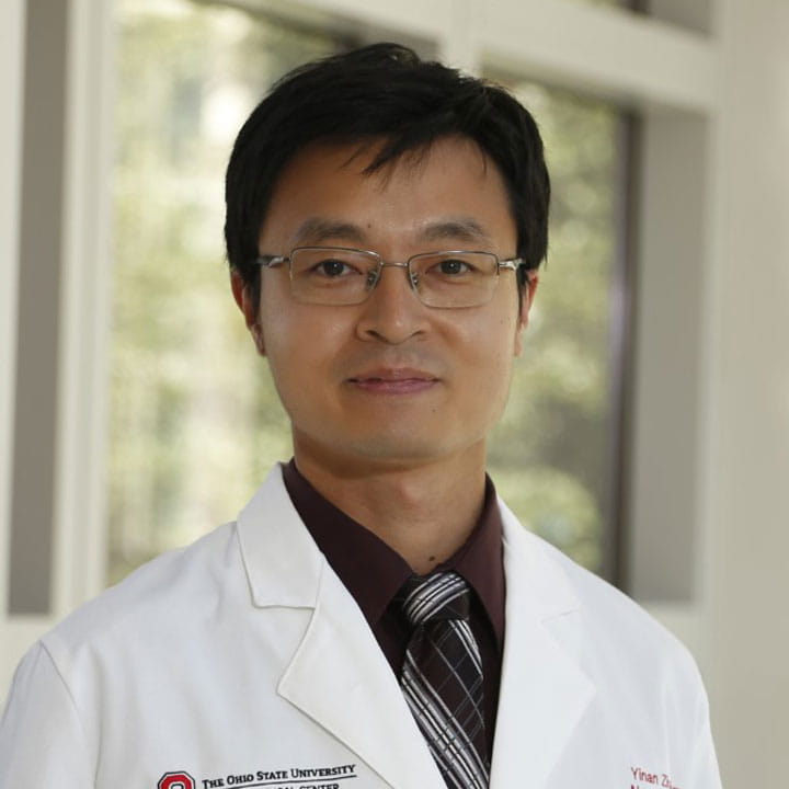
Yinan Zhang, MD
 Biological aging in people with multiple sclerosis
Age is the biggest driver of disease progression in multiple sclerosis. Biological age, which refers to the underlying cellular, molecular, and genetic processes of aging, may more accurately predict MS disease outcomes. Dr. Zhang's translational research studies serum markers of biological aging in people with MS that measure processes such as cellular senescence and epigenetic alterations. Our current findings show a subset of people with MS have accelerated biological aging that may be associated with progressive forms of MS and higher disability. Improved understanding of the role of biological aging in MS could lead to trials of existing and upcoming therapies that target aging processes as a new approach to treating MS disease progression.
Specific grants awarded: GEMSSTAR, K23
View Dr. Yinan Zhang's profile
Grace Shin
 Understanding peripheral mechanisms of pain to improve patient care for those suffering from cancer therapy-induced pain.
Treatments for cancer, such as chemotherapy and immunotherapy, are life-saving procedures for patients yet often result in unwanted side effects. In the US alone, more than 3 million cancer patients and survivors treated with chemotherapy suffer from a painful sensory disorder called chemotherapy-induced peripheral neuropathy (CIPN), with no effective clinical intervention. Our lab aims to comprehensively characterize the pathological progression leading to CIPN, with the goal of improving prediction, diagnosis, and prognosis for these patients.
Through a transdisciplinary approach, our work focuses on understanding the peripheral environment of pain-sensing neurons, specifically in the skin, where these neurons are surrounded by immune cells, keratinocytes, and fibroblasts. This very environment is where peripheral neuropathy starts, and neuronal plasticity in this area is proposed to contribute to the maintenance of neuropathic pain. However, we know very little about the inter- and intra-cellular changes in neurons and non-neuronal cells.
Our long-term goal is to significantly improve patient outcomes by translating fundamental findings into actionable immunomodulatory therapies for cancer therapy-induced pain while advancing the fields of sensory neuroscience and pain biology.
Specific grants awarded: K01 (PI), R21 (MPI), R01 (co-I), NSF (Collaborator)
View Dr. Grace Shin's profile View Shin lab
Luke Hammond
 Advanced light microscopy and quantitative techniques to enable and accelerate neuroscience research.
Mr. Hammond is the Director of Quantitative Imaging in the Department of Neurology. In this role, he collaborates with research teams to develop, apply, and disseminate advanced light microscopy and quantitative techniques to enable and accelerate neuroscience research. His work bridges the gap between advanced technology and neuroscience research, by creating innovative imaging and analysis solutions for complex research questions pertinent to neurodegenerative and neuroimmune conditions in the clinic.
He has developed open-source tools, including BrainJ and SpinalJ, which utilize machine learning for high-throughput analysis of whole mouse brains and spinal cords. These readily accessible tools allow automated organ reconstruction from tissue sections and accurate mesoscale mapping of cells and neuronal projections, which are essential for cell and circuit mapping in the CNS.
Recent work includes the development of Restoration Enhanced SPine and Neuron (RESPAN) analysis, a carefully validated application for accurate automated dendritic spine quantification. By integrating advanced imaging techniques with state-of-the-art quantification methods initially developed in basic neuroscience research, his work in the Department aims to provide comprehensive characterizations of neuron-immune dynamics in various pathological conditions, contributing to the development of novel therapies for neurological conditions.
Specific grants awarded: 2024-2029 - R01 Research Project Grant (PA-20-185), NINDS (Role: Co-investigator). “Role of immune cells in skin reinnervation by collateral sprouting after peripheral nerve injury.”
Luis Bonet-Ponce, PhD
 Lysosomes are central organelles to preserve cellular homeostasis, especially during stress. Their dysfunction is linked to neurodegenerative diseases. At the Bonet-Ponce lab, we study lysosomal damage to find therapeutic targets for these disorders.
The lysosome is key to cellular homeostasis and stress response. Dysfunctional lysosomes can lead to cell death and organ failure. Understanding lysosomal dynamics—movement, positioning, and maintenance—is crucial for grasping how cells adapt to internal and external changes.
Lysosomal dysfunction is linked to neurodegenerative diseases like Parkinson's disease, Alzheimer's disease, or Amyotrophic lateral sclerosis (ALS). In the Bonet-Ponce lab, we focus on studying lysosomal function during cellular stress. By studying lysosomal proteins and their mutations, we aim to understand their role in these diseases and explore potential therapies targeting lysosomal dysfunction.
View Luis Bonet-Ponce's profile Bonet-Ponce lab
